Saturday, April 13, 2013

Normal anatomy of the placenta & foetus

Normal anatomy of the placenta


Saggital projection

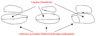
Placenta appears as an echogenic structure separated from the anechoic liquor in anterior or posterior walls of the uterus either in the upper segment of the fundus or in the lower segment closer to the cervical os or covering the os


Coronal projection
Placenta appears in lateral, anterior or posterior walls

Transducer on ant abdominal wall   
                 

Friday, April 12, 2013

Descriptive terms

Transducer

Echogenic lesions reflect ultrasound waves & appear as bright white echoes. eg.gas, calculi, calcifications
Transducer

Hypoechoic lesions partly transmits ultrasound waves appear as intermediate echoes eg.soft tissues, masses

Obstetric hemorrhage

Causes of obstetric hemorrhage

Ante partum
  • Ectopic pregnancy
  • Abortion
  • H mole
  • Placenta praevia
  • Placental abruption
Intra partum and post partum
  • Placenta praevia
  • Previous caesarian section with placenta praevia
  • Placental abruption
  • Placenta accrete
  • Uterine rupture
  • Uterine atony
  • Over distended uterus
  • Retained products of conception
  • Genital tract injury
  • Broad ligament tear

Diabetic foot ulceration

Background
  • Diabetes mellitus has reached epidemic proportions worldwide and its epicenter is in South Asia.
  • One of its main complications is lower extremity ulceration and infection leading to amputation. And this is the main contributor to lower limb amputations outside trauma. The good news is foot ulceration is preventable.
  • Furthermore aggressive appropriate management of early lesions prevents amputation.
  • This is achieved only with good knowledge and understanding of the pathophysiological processes involved.
  • This would be best achieved by teaching the subject as a stand-alone module.
  • It is important to remember that diabetics do not lose their limb overnight and 70-90% of amputations are preceded by ulceration.
  • It is a major contributor to amputation, ironically 30-50% of amputees lose the other limb within 5 yrs moreover approximately 100% die within 5 years of the second amputation.

Cardiopulmonary Resuscitation

Adult Cardiopulmonary Resuscitation                                       
Introduction
Cardio-respiratory arrest is cessation of mechanical activity of the heart. It is a clinical diagnosis evidenced by unresponsiveness, apnoea or agonal respirations and absence of a detectable central pulse. Immediate and systematic action is essential to prevent death or permanent cerebral damage.
Survival after cardiac arrest out of hospital is extremely low. Coronary artery disease is the commonest cause for sudden cardiac arrest, affecting nearly 700,000 people a year in Europe and one third of people developing a myocardial infarction die before reaching the hospital. The commonest presenting rhythm is rapid ventricular tachycardia or ventricular fibrillation. In the absence of immediate bystander initiated cardio-pulmonary resuscitation this deteriorates to asystole.
In- hospital cardiac arrest occurs in a sicker group of patients and only 20% of them will survive to leave the hospital. The presenting rhythm is either asystole or pulseless electrical activity.
Causes of cardiac arrest
Cardio-pulmonary arrest may occur due to airway obstruction, breathing inadequacy or cardiac abnormalities.
style1
Close