Sunday, September 8, 2013

Why the children are more prone to be dehydrated compared to adults.


  • Dehydrations reduction of body water level in the extracellular compartment sometimes accompanied with intracellular compartment due to excessive water loss or lack of water intake.
 
  • The signs of dehydration are;
      • Decrease skin turgor.
      • Dry lips & tongue.
      • Decrease blood pressure.
      • Increase heart rate & pulse .
      • Sunken eyeballs.
      • Sunken fontanelle (infants).
 
  • The symptoms of dehydration are;
      • Dry throat and mouth.
      • Difficulty in speech.
      • Decrease urine output.
      • Lethargy.
      • Weight loss.

Physiological basis of giving normal saline to a patient with isosmotic dehydration.


o   Dehydration is a decrease in ECF volume due to loss of H2O and solutes such as Na+.

o   In isoosmotic dehydration loss of H2O is equal to the loss of solutes.

o   Loss of isotonic fluid from plasma causes a loss of fluid from interstitium to plasma to maintain hydrostatic balance between the compartments.

o   But there is no change in the osmolality in the ECF compartment because the fluid lost is isotonic.

o   Thus there is no shift of fluid into or out of ICF because there is no change in ECF osmolality.

Up regulation and down regulation with examples

Down regulation


o   Is the process by which a cell decreases the quantity of a cellular component, such as RNA or protein in response to an external variable.

o   There are several ways in which a cell down regulates its cellular components.

1.      Receptor mediated endocytosis.

Occurs when the material to be transported into the cell binds to certain specific molecules in the membrane. Common cause for down regulation in membrane receptor. 

2.      Internalization.

Ligands bind to their receptors and ligand-receptor complex move laterally in the membrane to coated pits where they are taken into the cell by endocytosis. Some receptors are recycled and some replaced by de nova synthesis. 

The main characteristics of the erythrocyte cell membrane and relate these to the function of erythrocyte.

Organization of erythrocyte membrane


Human erythrocytes are small cells that lack nuclei when mature they appear as circular biconcave discs.
  • The membrane structure agrees with the Fluid Mosaic Model.
      • “the membrane protein ,intrinsic proteins(integral) deeply embedded and peripheral proteins loosely attached, that in an environment of fluid phospholipid bilayer.”
      • Fluidity of the membrane largely depends on the lipid composition of the membrane
      •  Due to presence of phospholipids erythrocyte membrane has high fluidity & flexibility.

The main sub cellular features of a protein secreting cell.


o   A protein secreting cell is metabolically very active
o   It receives signals via G protein coupled receptors.
o   It has a prominent nucleus which takes a large space of the cell. (Transcription, translation occurs in it.)
o   RER supports in synthesis of protein. So it is prominent in the cell. (RER has ribosome on its surface which involve in polypeptide synthesis using amino acids)
o   More mRNA, tRNA & rRNA make cytoplasm basophilic.
o   From RER synthesized polypeptides sent to Golgi apparatus for further modifications via vesicles. So large number of vesicles can be seen.
o   Well developed Golgi apparatus can be seen for post translational modifications of synthesized polypeptides.
o After that finished proteins exit from the cells by a secretory vesicles made out of Golgi apparatus, so large number of secretory vesicles can be seen.

The transport mechanism of monosaccharides across the intestinal mucosal cells.


o   Monosaccharides are formed by single saccharide molecules.
o   We normally talk about glucose, fructose & galactose as monosaccharides. There are so many except these three.
o   When we talk about starches, they are digested to various types of monosaccharides, disaccharides by gut enzymes secreted from various organs like pancreas.
o   There are same enzymes which digest disaccharides to monosaccharides. They are placed on brush border of the mucosal cells. Isomoltase, Moltase, Sucrose.
o   After the digestion final product is monosaccharides & they are transported across the intestinal mucosal cells to the portal system by various transport mechanisms.


How G protein help in signal transmission in to the cell from an incoming ligand & outline the steps.


  • Ligand bind G protein coupled receptor which has α ,β & γ subunits.
  • After binding, GTP in α dissociates from β & γ( all 3 subunits can produce physiological effects)
  • Causes production of a 2nd messenger eg:-cAMP via activation of adenylyl cyclase.
  • cAMP produce activated protein kinase A( pKA)  Ã   pKA phosphorylate certain molecules & activate /inhibit  à brings about the physiological action
  • When work is done , intrinsic GTPase activity of GTP-α converts GTP to GDP   à α ,β & γ subunits reassociates  à action terminates.

Monday, April 15, 2013

Management of Postpartum Collapse

COLLAPSE

Before delivery
 
After delivery
After 3rd stage
Rupture uterus
PPH – During 3rd stage
Ruptured uterus
Amniotic fluid embolism
 
Uterine inversion
 
RUPTURED UTERUS
History
  • Past caesarean section
  • Grand multigravida
  • Multigravida given oxytocin during labour
  • Almost never in primigravida - except following manipulations or previous uterine scar e.g. myomectomy

Management of Postpartum Collapse - AMNIOTIC FLUID EMBOLISM


Presentation
  • Extremely sudden severe shock with severe dyspnoea, cyanosis Pulmonary oedema
  • Blood stained frothy sputum
  • Convulsions may occur  
Onset
  • Usually immediately after membranes rupture however it can occur even with intact membranes occurs during strong uterine contractions
  • Usually fatal 

Saturday, April 13, 2013

Management of Postpartum Collapse - CONVULSIONS

Convulsions may be due to eclampsia, epilepsy, malaria, head injury, cerebral abscess, meningitis, encephalitis, etc.

Whatever the cause, initial steps are the same.
  • Stop the fits
  • Maintain airway
  • Prevent aspiration
  • Prevent injuries
Immediate care :
  • Position the patient - head low, turn to a side
  • airway - plastic or metal
  • Suck the mouth and pharynx
  • Place cushions around the patient or tie the limbs loosely with soft cloth  

Scan technique ii

Indications for 2nd & 3rd trimester scanning

  • Assigning dates
  • Assessing foetal growth, amniotic fluid volume, foetal heart motion & foetal presentation.
  • Assessing placental position & texture.
  • For review of foetal anatomy
  • To detect the presence of adenexal masses or cysts or uterine myomas
Scan technique
  • Bi parietal diameter measurement of the foetal head –BPD
  • BPD measurement is more accurate in the first trimester.
  • BPD is the distance between the anterior & posterior tables of the skull.
  • It is measured perpendicular to the mid line echo (falx)


Scan technique i

First trimester

Indications
  • Bleeding after a POA
  • Pain during pregnancy
  • Pregnancy complicated by fibroid or ovarian cyst
 
Preparation
Scans are performed though the full bladder using a 5 MHtz curvilinear probe.

Indications for first trimester sonography 
  • Localization of the GS –Intra uterine or Ectopic pregnancy
  • Identification of foetal viability
  • Anovular gestation
  • Estimation of POG
  • Assessment of multiple pregnancy, number of embryos chorionicity & amniocity

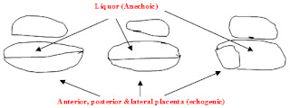
Normal anatomy of the placenta & foetus

Normal anatomy of the placenta


Saggital projection

Placenta appears as an echogenic structure separated from the anechoic liquor in anterior or posterior walls of the uterus either in the upper segment of the fundus or in the lower segment closer to the cervical os or covering the os


Coronal projection
Placenta appears in lateral, anterior or posterior walls

Transducer on ant abdominal wall   
                 

Friday, April 12, 2013

Descriptive terms

Transducer

Echogenic lesions reflect ultrasound waves & appear as bright white echoes. eg.gas, calculi, calcifications
Transducer

Hypoechoic lesions partly transmits ultrasound waves appear as intermediate echoes eg.soft tissues, masses

Obstetric hemorrhage

Causes of obstetric hemorrhage

Ante partum
  • Ectopic pregnancy
  • Abortion
  • H mole
  • Placenta praevia
  • Placental abruption
Intra partum and post partum
  • Placenta praevia
  • Previous caesarian section with placenta praevia
  • Placental abruption
  • Placenta accrete
  • Uterine rupture
  • Uterine atony
  • Over distended uterus
  • Retained products of conception
  • Genital tract injury
  • Broad ligament tear

Diabetic foot ulceration

Background
  • Diabetes mellitus has reached epidemic proportions worldwide and its epicenter is in South Asia.
  • One of its main complications is lower extremity ulceration and infection leading to amputation. And this is the main contributor to lower limb amputations outside trauma. The good news is foot ulceration is preventable.
  • Furthermore aggressive appropriate management of early lesions prevents amputation.
  • This is achieved only with good knowledge and understanding of the pathophysiological processes involved.
  • This would be best achieved by teaching the subject as a stand-alone module.
  • It is important to remember that diabetics do not lose their limb overnight and 70-90% of amputations are preceded by ulceration.
  • It is a major contributor to amputation, ironically 30-50% of amputees lose the other limb within 5 yrs moreover approximately 100% die within 5 years of the second amputation.

Cardiopulmonary Resuscitation

Adult Cardiopulmonary Resuscitation                                       
Introduction
Cardio-respiratory arrest is cessation of mechanical activity of the heart. It is a clinical diagnosis evidenced by unresponsiveness, apnoea or agonal respirations and absence of a detectable central pulse. Immediate and systematic action is essential to prevent death or permanent cerebral damage.
Survival after cardiac arrest out of hospital is extremely low. Coronary artery disease is the commonest cause for sudden cardiac arrest, affecting nearly 700,000 people a year in Europe and one third of people developing a myocardial infarction die before reaching the hospital. The commonest presenting rhythm is rapid ventricular tachycardia or ventricular fibrillation. In the absence of immediate bystander initiated cardio-pulmonary resuscitation this deteriorates to asystole.
In- hospital cardiac arrest occurs in a sicker group of patients and only 20% of them will survive to leave the hospital. The presenting rhythm is either asystole or pulseless electrical activity.
Causes of cardiac arrest
Cardio-pulmonary arrest may occur due to airway obstruction, breathing inadequacy or cardiac abnormalities.

Lumbar Puncture

INTRODUCTION
  • Lumbar puncture and analysis of cerebrospinal fluid is widely used as a diagnostic tool in neurological diseases.
  • Lumbar puncture (LP) is the insertion of a needle into the subarachnoid space in the lumbar region to obtain cerebrospinal fluid (CSF) for diagnostic or therapeutic purposes.
  • The CSF obtained by LP can provide crucial data in the diagnosis of life threatening conditions such as meningitis and encephalitis and evaluation of other disease conditions such as demyelinating diseases.
  • Lumbar puncture is alsoperformed for therapeutic reasons, such as the treatment of benign intracranial hypertension. 
  •  The spinal cord ends at the lower border of L1 vertebrae (Fig 1).
  • Below this are nerve roots – the cauda equina. Lumbar puncture can be safely done below L1-2 vertebral space, as there is no risk of injuring the spinal cord.

Management of a difficult airway and failed intubation

Introduction

  • Probably the most important job of an anaesthetist is to maintain a safe airway of an anaesthetised patient
  • Difficulty in airway management is the single most important cause of anaesthesia related morbidity and mortality     
  • If a difficult airway is detected and appropriate measures are taken to face it before anaesthetising a patient, morbidity and mortality associated with airway related problems can be minimised
  • Hence the importance of assessing the airway during the pre-operative visit
What is a Difficult Airway?

  • An airway which is difficult to be maintained or intubated or unable to be intubated in an anaesthetised or unconscious patient

IV Cannulation

Indications
  1. Administration of IV fluids
  2. Administration of blood products
  3. Administration of IV therapy
Procedure

  1. Confirm patient identity
  2. Communicate with the patient
    • Explain the procedure to the patient and get his/her verbal consent
    • Try to minimize the patient distress
    • Answer any questions the patient has
          
  3. Gather the equipment needed
    • Non sterile pair of gloves
    • Kidney tray
    • Tourniquet
    • Sterile alcohol swabs
    • IV Cannula of appropriate size
o Large bore cannulas are needed for rapid / thick fluid administration. E.g. 16 gauge gray canula
o The commonest used in day to day practice for IV fluid administration and drug administration are gauge 18 and 20

Wednesday, April 10, 2013

Anaphylaxis

Introduction-Anaphylactic reaction

· IgE -mediated type I hypersensitivity reaction.
· Effects are due to wide spread degranulation of mast cells.
· A dose independent unpredictable adverse drug reaction
 
Mechanism:
Commonly associated with a previous exposure to a similar group of drugs or chemicals. Foreign proteins act as antigens and stimulate production of IgEantibodies. They bind to mast cells and basophils, and with subsequent exposure massive degranulation occurs due to these already formed antibodies reacting with new antigens.
There is a massive release of mediators : Eg – Histamine, leukotrienes, tryptase, protease.
They give rise to bronchoconstriction, vasodilatation and increased capillary permeability. 

Malignant hyperthermia

Introduction.

  • Inherited disorder in skeletal muscle cells
  • Autosomal dominant
  • Variable penetrance
  • Incidence:-   1:15 000 – paediatrics         1: 40 000 – adult
  • Can be lethal
Aetiology.
  • A genetic mutation in Rynodine receptor, an intracellular calcium channel in sarcoplasmic reticulum.
  • When triggered, there is an exaggerated release of calcium ions causing hypermetabolism.
  • Triggering agents:- Halogenated inhalational anaesthetic agents Suxamethonium
  • They can be otherwise healthy individuals.
  • More susceptible in :-
            • Duchenne muscular dystrophy
            • Central core disease
            • Osteogenesis imperfecta
            • Strabismus surgery
            • Repeated anaesthetics
            • Positive family history.

Abdominal ultra sound

CHECK LIST ON ABDOMINAL ULTRA SOUND


Liver                    Size, margins, echogenecity, mass lesions, portal veins, hepatic veins
                       
Gall bladder         Size, calculi, sludge, cholecystitis

Bile ducts             Normal or dilated

Pancreas               Size, echogenecity, masses, calcification, duct dilatation

Spleen                  Size, mass lesions

Kidneys                Size, position, echo pattern, calculi, hydronephrosis, mass lesions

Oxygen therapy and Interpretation of blood gases

Oxygen therapy
  • Different modes of O2 therapy
-          nasal prongs
-          face mask
-          venturi mask
-          ET tube/LMA
  • Ordinary face mask provides a FiO2 of about 35%
  • Addition of an reservoir bag can increase the FiO2> 70% 
  • Venturi mask can provide a FiO2 up to about 60%
  • Anaesthetic circuit can provide a FiO2 close to 100%

Ventilation

Initiation of mechanical ventilation 

Objective of this station is to familiarize with clinical assessment of the patient before initiation of mechanical ventilation.

 Clinical parameters include:

  • Level of consciousness and patients ability to cooperate
  • Use of accessory muscles of respiration
  • Restlessness, anxiety, sweating and posture preferred by the patient
  • Presence of cyanosis
  • Altered chest wall movements
  • Pulse rate and rhythm disturbances
  • Oxygen saturation
  • Assess for effectiveness of cough reflex and phonation
  • Examination of lungs
Note: The decision to initiate mechanical ventilation should be made considering multiple parameters and not a single parameter. Especially normal saturation does not mean ventilation is normal. (Misnomer of most of clinicians).

Management of Shoulder Dystocia

The HELPERR Mnemonic


Basic and immediate intervention in management of shoulder dystocia.

H - Call for help.

This refers to activating the pre-arranged protocol or requesting the appropriate personnel to respond with necessary equipment to the labor and delivery unit.

E - Evaluate for episiotomy.

Episiotomy should be considered throughout the management of shoulder dystocia but is necessary only to make more room if rotation maneuvers are required. Shoulder dystocia is a bony impaction, so episiotomy alone will not release the shoulder. Because most cases of shoulder dystocia can be relieved with the Mc Roberts maneuver and supra pubic pressure, many women can be spared a surgical incision.

Electrocardiography (ECG)

Introduction

  • The ECG is a basic investigation that is widely used in medicine
  • You are guided through a module based on your objectives.
  • This package teaches you the important fundamentals of recording a 12-lead ECG and labeling the ECG.
  • Numerous patient cases are provided for evaluation while your understanding of material will be tested at the end of the module

Normal activation of the heart

  • The resting potential is maintained by ionic gradients across the cells with a higher sodium concentration outside and higher potassium concentration inside
  • Following a stimulus, a rapid inward movement of sodium occurs causing a loss of the negative internal potential. This is called as depolarization.
  • This is followed by repolarization, a slower process than depolarization where the potassium moves out of the cell

style1
Close