Monday, April 15, 2013

Management of Postpartum Collapse

COLLAPSE

Before delivery
 
After delivery
After 3rd stage
Rupture uterus
PPH – During 3rd stage
Ruptured uterus
Amniotic fluid embolism
 
Uterine inversion
 
RUPTURED UTERUS
History
  • Past caesarean section
  • Grand multigravida
  • Multigravida given oxytocin during labour
  • Almost never in primigravida - except following manipulations or previous uterine scar e.g. myomectomy

Management of Postpartum Collapse - AMNIOTIC FLUID EMBOLISM


Presentation
  • Extremely sudden severe shock with severe dyspnoea, cyanosis Pulmonary oedema
  • Blood stained frothy sputum
  • Convulsions may occur  
Onset
  • Usually immediately after membranes rupture however it can occur even with intact membranes occurs during strong uterine contractions
  • Usually fatal 

Saturday, April 13, 2013

Management of Postpartum Collapse - CONVULSIONS

Convulsions may be due to eclampsia, epilepsy, malaria, head injury, cerebral abscess, meningitis, encephalitis, etc.

Whatever the cause, initial steps are the same.
  • Stop the fits
  • Maintain airway
  • Prevent aspiration
  • Prevent injuries
Immediate care :
  • Position the patient - head low, turn to a side
  • airway - plastic or metal
  • Suck the mouth and pharynx
  • Place cushions around the patient or tie the limbs loosely with soft cloth  

Scan technique ii

Indications for 2nd & 3rd trimester scanning

  • Assigning dates
  • Assessing foetal growth, amniotic fluid volume, foetal heart motion & foetal presentation.
  • Assessing placental position & texture.
  • For review of foetal anatomy
  • To detect the presence of adenexal masses or cysts or uterine myomas
Scan technique
  • Bi parietal diameter measurement of the foetal head –BPD
  • BPD measurement is more accurate in the first trimester.
  • BPD is the distance between the anterior & posterior tables of the skull.
  • It is measured perpendicular to the mid line echo (falx)


Scan technique i

First trimester

Indications
  • Bleeding after a POA
  • Pain during pregnancy
  • Pregnancy complicated by fibroid or ovarian cyst
 
Preparation
Scans are performed though the full bladder using a 5 MHtz curvilinear probe.

Indications for first trimester sonography 
  • Localization of the GS –Intra uterine or Ectopic pregnancy
  • Identification of foetal viability
  • Anovular gestation
  • Estimation of POG
  • Assessment of multiple pregnancy, number of embryos chorionicity & amniocity

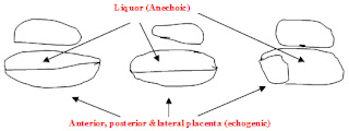
Normal anatomy of the placenta & foetus

Normal anatomy of the placenta


Saggital projection

Placenta appears as an echogenic structure separated from the anechoic liquor in anterior or posterior walls of the uterus either in the upper segment of the fundus or in the lower segment closer to the cervical os or covering the os


Coronal projection
Placenta appears in lateral, anterior or posterior walls

Transducer on ant abdominal wall   
                 

Friday, April 12, 2013

Descriptive terms

Transducer

Echogenic lesions reflect ultrasound waves & appear as bright white echoes. eg.gas, calculi, calcifications
Transducer

Hypoechoic lesions partly transmits ultrasound waves appear as intermediate echoes eg.soft tissues, masses

Obstetric hemorrhage

Causes of obstetric hemorrhage

Ante partum
  • Ectopic pregnancy
  • Abortion
  • H mole
  • Placenta praevia
  • Placental abruption
Intra partum and post partum
  • Placenta praevia
  • Previous caesarian section with placenta praevia
  • Placental abruption
  • Placenta accrete
  • Uterine rupture
  • Uterine atony
  • Over distended uterus
  • Retained products of conception
  • Genital tract injury
  • Broad ligament tear

Diabetic foot ulceration

Background
  • Diabetes mellitus has reached epidemic proportions worldwide and its epicenter is in South Asia.
  • One of its main complications is lower extremity ulceration and infection leading to amputation. And this is the main contributor to lower limb amputations outside trauma. The good news is foot ulceration is preventable.
  • Furthermore aggressive appropriate management of early lesions prevents amputation.
  • This is achieved only with good knowledge and understanding of the pathophysiological processes involved.
  • This would be best achieved by teaching the subject as a stand-alone module.
  • It is important to remember that diabetics do not lose their limb overnight and 70-90% of amputations are preceded by ulceration.
  • It is a major contributor to amputation, ironically 30-50% of amputees lose the other limb within 5 yrs moreover approximately 100% die within 5 years of the second amputation.

Cardiopulmonary Resuscitation

Adult Cardiopulmonary Resuscitation                                       
Introduction
Cardio-respiratory arrest is cessation of mechanical activity of the heart. It is a clinical diagnosis evidenced by unresponsiveness, apnoea or agonal respirations and absence of a detectable central pulse. Immediate and systematic action is essential to prevent death or permanent cerebral damage.
Survival after cardiac arrest out of hospital is extremely low. Coronary artery disease is the commonest cause for sudden cardiac arrest, affecting nearly 700,000 people a year in Europe and one third of people developing a myocardial infarction die before reaching the hospital. The commonest presenting rhythm is rapid ventricular tachycardia or ventricular fibrillation. In the absence of immediate bystander initiated cardio-pulmonary resuscitation this deteriorates to asystole.
In- hospital cardiac arrest occurs in a sicker group of patients and only 20% of them will survive to leave the hospital. The presenting rhythm is either asystole or pulseless electrical activity.
Causes of cardiac arrest
Cardio-pulmonary arrest may occur due to airway obstruction, breathing inadequacy or cardiac abnormalities.

Lumbar Puncture

INTRODUCTION
  • Lumbar puncture and analysis of cerebrospinal fluid is widely used as a diagnostic tool in neurological diseases.
  • Lumbar puncture (LP) is the insertion of a needle into the subarachnoid space in the lumbar region to obtain cerebrospinal fluid (CSF) for diagnostic or therapeutic purposes.
  • The CSF obtained by LP can provide crucial data in the diagnosis of life threatening conditions such as meningitis and encephalitis and evaluation of other disease conditions such as demyelinating diseases.
  • Lumbar puncture is alsoperformed for therapeutic reasons, such as the treatment of benign intracranial hypertension. 
  •  The spinal cord ends at the lower border of L1 vertebrae (Fig 1).
  • Below this are nerve roots – the cauda equina. Lumbar puncture can be safely done below L1-2 vertebral space, as there is no risk of injuring the spinal cord.

Management of a difficult airway and failed intubation

Introduction

  • Probably the most important job of an anaesthetist is to maintain a safe airway of an anaesthetised patient
  • Difficulty in airway management is the single most important cause of anaesthesia related morbidity and mortality     
  • If a difficult airway is detected and appropriate measures are taken to face it before anaesthetising a patient, morbidity and mortality associated with airway related problems can be minimised
  • Hence the importance of assessing the airway during the pre-operative visit
What is a Difficult Airway?

  • An airway which is difficult to be maintained or intubated or unable to be intubated in an anaesthetised or unconscious patient

IV Cannulation

Indications
  1. Administration of IV fluids
  2. Administration of blood products
  3. Administration of IV therapy
Procedure

  1. Confirm patient identity
  2. Communicate with the patient
    • Explain the procedure to the patient and get his/her verbal consent
    • Try to minimize the patient distress
    • Answer any questions the patient has
          
  3. Gather the equipment needed
    • Non sterile pair of gloves
    • Kidney tray
    • Tourniquet
    • Sterile alcohol swabs
    • IV Cannula of appropriate size
o Large bore cannulas are needed for rapid / thick fluid administration. E.g. 16 gauge gray canula
o The commonest used in day to day practice for IV fluid administration and drug administration are gauge 18 and 20

Wednesday, April 10, 2013

Anaphylaxis

Introduction-Anaphylactic reaction

· IgE -mediated type I hypersensitivity reaction.
· Effects are due to wide spread degranulation of mast cells.
· A dose independent unpredictable adverse drug reaction
 
Mechanism:
Commonly associated with a previous exposure to a similar group of drugs or chemicals. Foreign proteins act as antigens and stimulate production of IgEantibodies. They bind to mast cells and basophils, and with subsequent exposure massive degranulation occurs due to these already formed antibodies reacting with new antigens.
There is a massive release of mediators : Eg – Histamine, leukotrienes, tryptase, protease.
They give rise to bronchoconstriction, vasodilatation and increased capillary permeability. 

Malignant hyperthermia

Introduction.

  • Inherited disorder in skeletal muscle cells
  • Autosomal dominant
  • Variable penetrance
  • Incidence:-   1:15 000 – paediatrics         1: 40 000 – adult
  • Can be lethal
Aetiology.
  • A genetic mutation in Rynodine receptor, an intracellular calcium channel in sarcoplasmic reticulum.
  • When triggered, there is an exaggerated release of calcium ions causing hypermetabolism.
  • Triggering agents:- Halogenated inhalational anaesthetic agents Suxamethonium
  • They can be otherwise healthy individuals.
  • More susceptible in :-
            • Duchenne muscular dystrophy
            • Central core disease
            • Osteogenesis imperfecta
            • Strabismus surgery
            • Repeated anaesthetics
            • Positive family history.

Abdominal ultra sound

CHECK LIST ON ABDOMINAL ULTRA SOUND


Liver                    Size, margins, echogenecity, mass lesions, portal veins, hepatic veins
                       
Gall bladder         Size, calculi, sludge, cholecystitis

Bile ducts             Normal or dilated

Pancreas               Size, echogenecity, masses, calcification, duct dilatation

Spleen                  Size, mass lesions

Kidneys                Size, position, echo pattern, calculi, hydronephrosis, mass lesions

Oxygen therapy and Interpretation of blood gases

Oxygen therapy
  • Different modes of O2 therapy
-          nasal prongs
-          face mask
-          venturi mask
-          ET tube/LMA
  • Ordinary face mask provides a FiO2 of about 35%
  • Addition of an reservoir bag can increase the FiO2> 70% 
  • Venturi mask can provide a FiO2 up to about 60%
  • Anaesthetic circuit can provide a FiO2 close to 100%

Ventilation

Initiation of mechanical ventilation 

Objective of this station is to familiarize with clinical assessment of the patient before initiation of mechanical ventilation.

 Clinical parameters include:

  • Level of consciousness and patients ability to cooperate
  • Use of accessory muscles of respiration
  • Restlessness, anxiety, sweating and posture preferred by the patient
  • Presence of cyanosis
  • Altered chest wall movements
  • Pulse rate and rhythm disturbances
  • Oxygen saturation
  • Assess for effectiveness of cough reflex and phonation
  • Examination of lungs
Note: The decision to initiate mechanical ventilation should be made considering multiple parameters and not a single parameter. Especially normal saturation does not mean ventilation is normal. (Misnomer of most of clinicians).

Management of Shoulder Dystocia

The HELPERR Mnemonic


Basic and immediate intervention in management of shoulder dystocia.

H - Call for help.

This refers to activating the pre-arranged protocol or requesting the appropriate personnel to respond with necessary equipment to the labor and delivery unit.

E - Evaluate for episiotomy.

Episiotomy should be considered throughout the management of shoulder dystocia but is necessary only to make more room if rotation maneuvers are required. Shoulder dystocia is a bony impaction, so episiotomy alone will not release the shoulder. Because most cases of shoulder dystocia can be relieved with the Mc Roberts maneuver and supra pubic pressure, many women can be spared a surgical incision.

Electrocardiography (ECG)

Introduction

  • The ECG is a basic investigation that is widely used in medicine
  • You are guided through a module based on your objectives.
  • This package teaches you the important fundamentals of recording a 12-lead ECG and labeling the ECG.
  • Numerous patient cases are provided for evaluation while your understanding of material will be tested at the end of the module

Normal activation of the heart

  • The resting potential is maintained by ionic gradients across the cells with a higher sodium concentration outside and higher potassium concentration inside
  • Following a stimulus, a rapid inward movement of sodium occurs causing a loss of the negative internal potential. This is called as depolarization.
  • This is followed by repolarization, a slower process than depolarization where the potassium moves out of the cell

Tuesday, April 9, 2013

Disinfection and sterilization

Importance of sterilization,
In health care setting it is important;
  • To render articles and environment safe from the risk of transmitting infections.
  •  Pathogens on hands & environment are removed or destroyed by ,
        high standard of cleanliness
        drying, washing, good ventilation and sun light.
 
o   Wards should be well ventilated and sunny.

● High risk procedures

o       Require absolute sterility

        Surgery
        preparation

         of culture media
         Iv fluids and other injectable drugs

        Types of research work

Sharp tools

●   Sharp tools in use in hospital setting:
      • Syringes and needles
      • Scalpel blades
      • Razors
      • Infusion sets
      • Lancets
      • Broken glass
Why proper handling and disposal of sharps is important?
  • Improper handling and disposal of sharps leads to acquisition of infections by Healthcare Workers during their work ( occupational health hazards).
  • This leads to transmission of blood borne infections namely HBV, HCV AND HIV  infections among patients as well as Healthcare workers.
  • Proper handling and disposal of sharps leads to the minimization of the risk of getting infected by above blood borne infections. 
 

Hand hygiene

  • On average, 8.7% of hospital patients suffer nosocomial infections.
  • Hospital infections are a leading cause of death in some parts of the world.
  • Most patient deaths and suffering attributable to health care-associated infections can be prevented.
  • The costs of health care-associated infections vary by country and institution. 
  • The single most important measure to prevent and control health care -associated.
Benefits of hand washing are,
    • Reduces nosocomial infections
    • Preventing cross transmission of multi-resistant organisms
    • Preventing and control epidemics
    • Cost saving
    • Reduces morbidity and mortality

A guide to performing a bone marrow aspiration


1. Decide on the most appropriate site for aspiration;
  • The usual site for puncture in adults is the posterior superior iliac spine (PSIS).
  • This may be technically difficult in obese or immobile subjects.
  • The main advantage of this site is that a trephine biopsy can also be performed. Alternatively the sternum ( manubrium or 1st or 2nd parts of the body) can be used with care as there is a danger of  perforating the inner cortical layer and damaging the underlying large blood vessels and right atrium with serious consequences.
2. Obtain informed written consent;
  • The procedure (in brief) and its main adverse effects (although rare) should be explained to the patient prior to obtaining consent.
  • Pain, bleeding/haematoma formation and infection at aspiration site are the most commonly seen adverse effects, however, very rarely cause major concern.
3. Positioning the patient;
  • Either left or right lateral decubitous on a bed/couch, with the dorsal surface of the patient facing the operator.
  • Hold the hips and knees flexed to a comfortable maximum – knees drawn up and tucked into chest.

Venepuncture

Introduction Venepuncture is the term used for the procedure of entering a vein with a needle.

Indications
  1. Withdrawal of blood for investigations
  2. As a route for venous access
Procedure
  1. Confirm patient identity   
  2. Communicate with the patient
    • Explain the procedure to the patient and get his/her verbal consent
    • Try to minimize the patient distress
    • Ask if blood has been withdrawn previously
    • Sometimes the patient is able to point out sites where blood can be withdrawn successfully.
    • Answer any questions the patient has  
  3. Gather the equipment needed
    • Non sterile pair of gloves
    • Kidney tray
    • Tourniquet
    • Sterile alcohol swabs
    • Sterile needle and syringe of an appropriate size
    • Plaster / hypoallergenic tape
    • Blood collecting tubes or bottles
    • Ensure there is adequate light  

Technique of interpretation of plain radiograph of chest

CHECK LIST FOR PA FILM


Study the request form,
  • Patient’s age sex, clinical information.
  • Check serial number of radiograph against number on request form.
Technical aspects     
  • Correct position of patient
  • Area of interest - lung apices,lower neck, diaphragm, lateral chest walls. 
  • Exposure factors.   

Chemical agents used in sterilization and disinfection


Liquids
Agent
Purpose ,  Antiseptic & Disinfectant activity,
1.    Alcohols
70% ethanol,
70% isopropyl alcohol
(Up to 95% can be used but not in the pure form)
Kills – most vegetative bacteria, fungi & viruses
Inactive against spores of bacteria & fungi
1.    Skin disinfectant for venepuncture, cannula insertion & IM injections.
2.    Disinfection of thermometers and surfaces.
3.    Can be used as a base for other bactericides.
eg: Chlorhexidine, Iodine, Triclosan -  For pre-operation skin disinfection
-  eg: 0.5% chlorhexidine in 95%  ethanol
 
2.    Aldehydes
*Gluteraldehyde (Cidex)
2% gluteraldehyde is very commonly used in hospitals.


Kills – all organisms & spores
1.    2% Gluteraldehyde is used to disinfect heat sensitive instruments
eg. Fiber optic endoscopes , Dialysis equipment,  Plastic tubing.
1.    Gluteraldehyde – inactivated less by organic matter.
2.    Need proper cleaning of instruments before dipping.
3.    Tubes must be totally immersed & completely filed.
4.    Non corrosive
5.    Irritant to skin, eyes & mucous membrane – need proper washing with sterile water after immersion.
6.    Acidic solutions are slower sporicides than alkaline solutions.
7.    Should be used within 14 days or discarded following 20 immersions even before 14 days.
*Formaldehyde
Decontaminate postmortem rooms, & laboratories.(fumigation)
 

Monday, April 8, 2013

Tendon Suture

Tendons are relatively avascular structures and heal by the in growth of connective tissue from the epitenon.

Indications

  • traumatic injury to tendons
  • as part of another procedure
Assessment

    • Wounds in the vicinity of a tendon, assume tendon injury until it is shown to be intact on clinical examination
    • If no action is demonstrated and there is doubt, explore the wound 


style1
Close